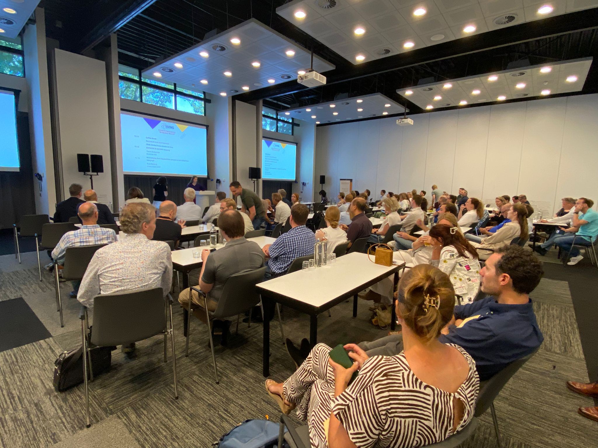
Met een volle zaal werd de wetenschappelijke voorjaarsbijeenkomst van de NVNG op 13 juni 2025 in het Postillion hotel te Bunnik gehouden met als thema Do you have the guts. Na de welkomstwoorden van de voorzitter van de Commissie Wetenschappelijke Ontmoetingen, (CWO) dr. Tineke van de Weijer, startte de eerste ochtendsessie It’s all about the Guts van het door de CWO samengestelde programma onder voorzitterschap van dr. Tineke van de Weijer en dr. Erik Aarntzen met een presentatie van nucleair geneeskundige drs. Peter Kaldeway (Antonius Ziekenhuis Nieuwegein) over Gastric Emptying, an update of the old protocols. Vervolgens behandelde klinisch radiochemicus dr. Gert Luurtsema (UMCG) het onderwerp Oral intake of PET-tracers. Tenslotte heeft gastroenterologist dr. Rianne Beckers (MUMC) Gastro-intestinal bleeding from diagnosis to treatment gepresenteerd.
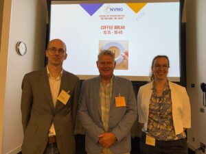
Sprekers van eerste ochtendsessie met van links naar rechts Peter Kaldeway, Gert Luurtsema en Rianne Beckers.
Na de pauze startte de tweede ochtendsessie Liver and Pancreas onder voorzitterschap van dr. Hendrikus Boersma en drs. Anke de Vries met een presentatie van nucleair geneeskundige dr. Tessa Brabander (Erasmus MC) betreffende DOTATOC in GEPNET imaging. Vervolgens behandelde nucleair geneeskundige prof. dr. Roel Bennink (Amsterdam UMC, tegenwoordig LUMC) het onderwerp Mebrofenin, from hepatobiliary atrophy to functional liver. De sessie werd voorgezet met de presentatie van nucleair geneeskundige prof. dr. Lioe-Fee de Geus-Oei, The value of [18F]FDG imaging in the treatment evaluation of colorectal liver metastasis, en vervolgens met Shit in, shit out; Internal Contamination door dr. Robert Westland (Amsterdam UMC).
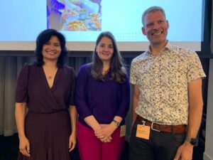 Sprekers van de tweede ochtendsessie met van links naar rechts Lioe-Fee de Geus-Oei, Tessa Brabander en Robert Westland (Roel Bennink niet op de foto).
Na de lunchpauze werd in de middag een sessie gehouden onder voorzitterschap van dr. Anke de Vries en dr. Maurits Wondergem. Voorafgaand aan de te presenteren abstracts ontvingen dr. Lucas Mangnus (NKI/AVL) en dr. Sanne Jansen (NKI/AVL) hun fellowshipcertificaten bij het afronden van hun stages.
In aansluiting op de fellowshipceremonie werden vier vrije inzendingen gepresenteerd. Onderzoeker Fleur Kleiburg (LUMC) presenteerde PSMA expression and PSMA PET/CT imaging in metastatic soft tissue sarcoma patients, results of a prospective study. Vervolgens presenteerde H. Schouw (UMC Groningen) In-vitro and ex-vivo targeted fluorescence imaging of the calcium-sensing receptor with fluorescein and 800CW coupled cinacalcet, gevolgd door nucleair geneeskundige Jouke Boer (Spaarne Gasthuis) met het onderwerp Optimizing the Coronary Care Pathway: Evaluating the Diagnostic Value of Combined CCTA and PET Imaging in a Rapid Access Clinic. De laatste spreker van de abstract sessie werd onderzoeker Mara Veenstra (ErasmusUMC) met The effect of [18F]F-FAPI PET/CT on management in patients with potentially resectable biliary tract cancers: a prospective multicentre study and cost-effectiveness analysis.
De samenvattingen van deze presentaties zijn te lezen aan het eind van dit verslag.
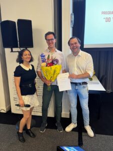
Lucas Mangnus (foto links) en Sanne Jansen (foto rechts) tussen Emilia Owers en Arthur Braat ontvangen fellowschipcertificaten en bloemen.
 Sprekers van de eerste middagsessie met van links naar rechts Fleur Kleiburg, , Jouke Boer en Mara Veenstra (H. Schouw niet op de foto).
Na de pauze werd de sessie Abdominal oncology – what’s new? gehouden onder voorzitterschap van dr. Hendrikus Boersma en dr. Maurits Wondergem, met als gastsprekers nucleair geneeskundige Ayça Arçay Öztürk (Institut Jules Bordet / Hôpital Universitaire de Bruxelles)) met het onderwerp FAPI and peritonitis carcinomatosa, en chirurg prof. dr. Go van Dam (Tracer BV) met de presentatie Instestinal imaging of oncology and inflammation, a clinical perspective. De laatste spreker van de dag was klinisch fysicus prof. dr. Ronald Boellaard (Amsterdam UMC) met Abdominal dynamic FDG-PET; what the FUR?.
 Sprekers van de slotsessie van het symposium met van links naar rechts Ayça Arçay Öztürk, Go van Dam en Ronald Boellaard.
Samenvattingen vrije inzendingen middagprogramma
PSMA expression and PSMA PET/CT imaging in metastatic soft tissue sarcoma patients, results of a prospective study
- Kleiburg1,2,, T. van der Hulle3, H. Gelderblom3, M. Slingerland3, F.M. Speetjens3, L.J.A.C. Hawinkels4, P. Dibbets-Schneider2, F.H.P. van Velden2, M. Pool5, S.W. Lam6, J.V.M.G. Bovée6, L. Heijmen2, and L.F. de Geus-Oei1,2,7
1Biomedical Photonic Imaging Group, University of Twente, Enschede, 2Department of Radiology, Section of Nuclear Medicine, 3Department of Medical Oncology, 4Department of Gastroenterology,5Department of Clinical Pharmacy and Toxicology, and 6Department of Pathology, Leiden University Medical Centre, 7Department of Radiation Science and Technology, Technical University of Delft.
Aim
Prostate-specific membrane antigen (PSMA) expression has been observed in a subset of soft tissue sarcomas, mainly in the neovascular endothelial cells. This feasibility study aimed to evaluate PSMA expression and PSMA PET/CT imaging in metastatic soft tissue sarcoma, providing important insights for potential future exploration of PSMA-targeted radioligand therapy.
Methods
This prospective single-centre study included adult patients with metastatic soft tissue sarcoma, with measurable disease (lesion diameter > 1 cm), available biopsy/resection material, ECOG/WHO performance status of 0-2 and either no prior systemic treatment, progressive disease during/after treatment, or stable disease/partial response with the last dose > 8 weeks prior. Immunohistochemical PSMA staining was performed on previously obtained biopsy or resection material. In case of high PSMA expression, as defined in previous literature, an [18F]-JK-PSMA-7 PET/CT scan evaluated tracer uptake, with adequate uptake defined as SUVmax > 8.
Results
Of 25 included patients, 11 (44%) had high PSMA expression: 4/11 leiomyosarcomas, 3/4 dedifferentiated liposarcomas, 2/5 undifferentiated pleomorphic sarcomas, 1/2 myxofibrosarcomas and 1/1 malignant peripheral nerve sheath tumour. Five of eleven patients agreed to an [18F]-JK-PSMA-7 PET/CT, of which three had lesions that showed adequate tracer uptake (SUVmax 10.7-16.7). However, uptake across all metastatic lesions was highly heterogeneous (median SUVmax = 3.8; range 0.5-16.7), indicating that these patients are unlikely to benefit sufficiently from PSMA-targeted therapy. The study was therefore terminated prematurely.
Conclusion
PSMA expression and PSMA tracer uptake in metastatic soft tissue sarcoma were highly heterogeneous. A deeper understanding of PSMA biology and improved patient selection criteria are essential for future application of PSMA-targeted radioligand therapy in this disease.
Trial registration: clinicaltrials.gov, NCT05522257. Registered 31-08-2022.
Optimizing the Coronary Care Pathway: Evaluating the Diagnostic Value of Combined CCTA and PET Imaging in a Rapid Access Clinic
J.J. Boer1, B. Lommen1, P. Bot2
Department of Nuclear Medicine1 and Cardiology2, Spaarne Gasthuis, Haarlem.
Introduction
To improve the efficiency and diagnostic accuracy of coronary artery disease (CAD) evaluation, the cardiology street (a rapid-access outpatient diagnostic pathway) at Spaarne Gasthuis has introduced a combined diagnostic approach using coronary computed tomography angiography (CCTA) and myocardial perfusion PET/CT. The diagnostic process is initiated with a coronary calcium score (CCS); patients with a CCS <250 proceed to CCTA, while those with higher scores or unfavourable plaque distribution undergo stress-only PET imaging. Additionally, PET is used to assess functional relevance in patients with >50% soft plaque stenosis on CCTA. This study investigates whether smarter decision-making based on the combination of CCTA and PET can reduce unnecessary invasive testing and optimize the care pathway.
Methods
A retrospective cross-sectional analysis was performed in 171 patients referred to the cardiology street. Data on CCS, CCTA, PET imaging results, and subsequent management decisions were collected. The pretest likelihood for CAD was assessed to evaluate adherence to the protocol (i.e., inclusion of patients with <50% pretest likelihood). The main outcome was the proportion of patients in whom combined non-invasive imaging could replace invasive coronary angiography (CAG).
Results
Relevant CAD was excluded in 87.1% of patients, with CCTA alone sufficient in 63.8% and PET in 21.5%. In six patients, both PET and CCTA fully matched the CAG result, rendering the invasive procedure redundant. Despite protocol criteria, 15% of patients underwent CAG, often without added value. Overdiagnosis was minimal, with only 5.7% of CCTA scans considered unnecessary. In total, combined CCTA and PET imaging enabled accurate diagnosis and treatment planning in 80% of patients without the need for CAG.
Conclusion
The combination of CCTA and PET imaging offers high diagnostic accuracy and reduces the need for invasive CAG in patients evaluated in a rapid diagnostic clinic. This strategy supports smarter, individualized diagnostic pathways that minimize overdiagnosis and unnecessary testing. Further studies should evaluate the scalability of this model and its impact on healthcare efficiency.
FAPIChol trial: The effect of [18F]F-FAPI PET/CT on management in patients with potentially resectable biliary tract cancers: a prospective multicentre study and cost-effectiveness analysis.
EUCT 2023-507938-24-00; NCT06355427
M.M.K. Veenstra1, E. Vegt1, B. Groot Koerkamp2, M. Lam3, J. Hagendoorn4, R. Bennink5, R.J. Swijnenburg6, F. A. Verburg1, M.G.J. Thomeer1
1Department of Radiology and Nuclear Medicine, 2Department of Surgery, Erasmus MC University Medical Centre Rotterdam, 3Department of Radiology and Nuclear Medicine, 4Department of Surgery, Regional Academic Cancer Centre Utrecht (RAKU), University Medical Centre Utrecht, 5 Department of Radiology and Nuclear Medicine, 6Department of Surgery, Amsterdam University Medical Centres.
Rationale
Cholangiocarcinoma is a rare but aggressive hepatobiliary cancer, with surgical resection as the only curative treatment. However, up to 29% of patients initially deemed resectable are found to have metastases during surgery. Current imaging (CT, MRI, and FDG PET/CT) lacks sufficient accuracy to reliably detect metastatic disease preoperatively. Fibroblast activation protein inhibitor (FAPI) PET/CT has shown promise in detecting metastases in desmoplastic tumours like cholangiocarcinoma, with superior performance over FDG PET/CT. This study aims to determine whether incorporating [18F]F-FAPI PET/CT into preoperative assessment improves staging accuracy, prevents futile surgeries, and is cost-effective.
Study goals and objectives
This study assesses the diagnostic accuracy of [18F]F-FAPI PET/CT in detecting metastases in patients with potentially resectable proximal cholangiocarcinoma. Additionally, it evaluates whether integrating [18F]F-FAPI PET/CT alters treatment plans and proves cost-effective.
Primary endpoint
- diagnostic accuracy (sensitivity, specificity, PPV, and NPV) of [18F]F-FAPI PET/CT
Secondary endpoints
- per-region and per-lesion diagnostic accuracy
- impact on treatment decisions
- cost-effectiveness
- additional significant findings
- inter-reader variability
- diagnostic delay
- incidence/severity of AEs/SAEs (CTCAEv5)
- improvement of accuracy by distinguishing benign/malignant disease using dynamic [18F]F-FAPI PET/CT (subgroup analysis, n = 30)
Trial population
Adults (≥18 years) with potentially resectable proximal cholangiocarcinoma (perihilar, intrahepatic, gallbladder) eligible for surgery based on CT/MRI. Exclusion criteria: prior abdominal surgery/chemotherapy, pregnancy/lactation, or FDG PET/CT indication.
Total sample size: n = 81.
Interventions
If patients are deemed eligible for surgery by the weekly organ-specific multidisciplinary team, patients will be asked to participate in the study. After inclusion, patients will receive [18F]F-FAPI PET/CT, followed by six months follow-up. Follow-up includes the collection of data about additional imaging, surgery, other treatments or complications. To determine cost-effectiveness, patients’ medical consumption outside the hospital will be elicited using two questionnaires. Patients will be asked to fill out the ‘institute for Medical Technology Assessment (iMTA) Medical Consumption Questionnaire (iMCQ)’ at three and six months after [18F]F-FAPI PET/CT. Patients will also be asked to fill out the European Organisation for Research and Treatment of Cancer (EORTC) group’s QLQ-C30 and QLQ BIL21 questionnaires and the EuroQol (EQ) Group’s EQ-5D-5L questionnaire once every two weeks during the first two months after [18F]F-FAPI PET/CT and then once every four weeks until six months follow-up.
In-vitro and ex-vivo targeted fluorescence imaging of the calcium-sensing receptor with fluorescein and 800CW coupled cinacalcet.
H.M. Schouw1,2,5, A.J.M. Korenhof3,6, M. Kozanhan3,6, M.E. Noltes1,2, F. Reessing4, C.L.F. van Beek7, A.H. Brouwers1, S Kruijff1,2,5, W. Szymanski4,7,8, A.P. NagelkerkB3,6, H.H. Boersma1,3
1 Department of Nuclear Medicine and Molecular Imaging, 2Department of Surgery, 3Department of Clinical Pharmacy and Pharmacology, 4Department of Radiology, Medical Imaging Center, University Medical Centre Groningen, 5Department of Molecular Medicine and Surgery, Karolinska Institutet, Stockholm, Sweden, 6Pharmaceutical Analysis, Groningen Research Institute of Pharmacy, Faculty of Science and Engineering, 7Center for Systems Chemistry, Stratingh Institute for Chemistry, 8Department of Medicinal Chemistry, Photopharmacology and Imaging, Groningen Research Institute of Pharmacy, University of Groningen.
Aim
The parathyroid glands are four small structures adjacent to the thyroid gland that regulate calcium homeostasis through parathyroid hormone secretion and calcium-sensing receptor (CaSR) expression. Inadequate identification during surgery can result in (permanent) hypoparathyroidism with possible symptoms of muscle cramps, seizures, fatigue and depression. Fluorescently labelled tracers offer a promising approach for enhancing intraoperative visualization, potentially improving surgical precision to spare vascular structures and parathyroid perfusion. We developed a cell-based CaSR imaging model to investigate the feasibility of this concept.
Materials and Methods
A CaSR-targeting fluorescent tracer was developed by conjugating the calcimimetic drug cinacalcet to either fluorescein (λex 495 nm, λem 520 nm) or IRDye800CW (λex 775 nm, λem 792 nm). Different cell lines expressing CaSR, including the COLO320 cell line, were employed. Binding of the fluorescent cinacalcet tracers at various concentrations was assessed using fluorescence microscopy, flow cytometry, and plate reader assays. At this moment, possibilities are explored to use 18F-cinacalcet as a control tracer. Lastly, fresh parathyroid adenoma tissue was incubated post-operatively to evaluate ex-vivo binding of the fluorescent tracers to CaSR.
Results
Incubation of COLO320 cells with increasing concentrations of cinacalcet-fluorescein, displayed increasing levels of fluorescence, as measured by flow cytometry. Appearance of fluorescence was also time-dependent and reduced when unlabelled cinacalcet was added. These results were further established with cinacalcet-800CW, as well as with fluorescence microscopy and spectrophotometry measurements. Currently, we are confirming the results obtained in dedicated genetically modified COLO320 cells, engineered to either overexpress or silence CaSR expression. Incubation of fresh parathyroid adenoma tissue with both tracers showed visible retention of tissue fluorescence after extensive rinsing, further validating suitability of the developed tracers.
Conclusion
This study presents the successful synthesis of CaSR-targeting fluorescent tracers, cinacalcet-fluorescein and cinacalcet-800CW, which exhibit binding to the CaSR receptor in in-vitro and ex-vivo assays. These tracers demonstrate significant potential for enhancing CaSR imaging, enabling more accurate visualization of the parathyroid gland during surgery. This advancement offers a promising translational tool for the clinical improvement of surgical precision and hopefully reducing post-operative hypoparathyroidism.